Optical Coherence Tomography (OCT) Imaging
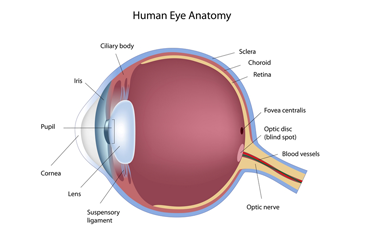
Optical Coherence Tomography (OCT) uses an infrared light to image the central part of the retina called the macula. These images represent a virtual cross-section of the retina, allowing the ophthalmologist to see microscopic details of the macula. OCT is useful for diagnosing, treating, and following several retinal diseases, including age related macular degeneration, diabetic retinopathy, vein occlusion, and macular holes and macular puckers. An OCT is frequently performed in the clinic after your eyes have been dilated, and takes only minutes to complete.

