
Macular Degeneration Specialists in Sarasota and Bradenton
Early diagnosis and consistent treatment can help preserve your central vision.
Age-related macular degeneration affects the part of the retina responsible for reading, driving, and recognizing faces. Our retina specialists use advanced imaging and proven treatments to detect changes early, slow disease progression, and help you maintain the best vision possible.
If you are over 60 or have a family history of A.M.D., regular retinal exams are one of the most effective steps you can take to protect your sight.
Shane Retina > Patient Services & Education > Macular Degeneration
At a Glance: Macular Degeneration
Age-related macular degeneration (A.M.D.) is a breakdown of the macula, the small center of the retina that gives you sharp central vision for reading, driving, and recognizing faces.
- Who gets it: Mostly people over 60. Risk rises with age, family history, and smoking.
- Two forms: Dry A.M.D. (about 90% of patients) usually progresses slowly. Wet A.M.D. (about 10%) can change vision quickly and is treatable with eye injections.
- What it does: Blurs or distorts central vision. Side vision is usually preserved, and macular degeneration rarely causes total blindness.
- What to do now: Sudden distortion, a new blind spot, or a quick drop in vision in one eye should be evaluated the same day. Call (941) 351-1200.
- Otherwise: Schedule a dilated retinal exam. Early diagnosis gives you the most treatment options.
Shane Retina treats macular degeneration at two Sarasota offices and in Venice.
Medically reviewed by Thomas Shane, M.D., Retina Specialist – Last updated September 4th, 2026
Age-related vision changes are to be expected; they happen throughout our entire lives, and in our later years they tend to progress more rapidly. While routine changes are perfectly normal, as we get older we must be cognizant of the potential for age-related macular degeneration, or A.M.D.
What is the Macula?
The origin of the word macula can be found in Latin, where it was taken to mean ‘spot.’ In regards to the eye, the macula refers to the central 5 millimeters of the retina, where vision is the sharpest. The center of the macula does have a central ‘spot’ in fact, called the fovea. Your entire ability to read and drive is located within this critical one millimeter of the central retina.
There are many conditions that can affect the macula. While macular degeneration is the most common, you may also hear about other retinal problems such as macular pucker, macular hole, and macular dystrophy. Each of these conditions is unique, with distinct treatments and prognosis for vision.
What is Age-Related Macular Degeneration?
Age-related macular degeneration is a deterioration of the central retina that leads to a loss of visual acuity. Macular degeneration is described as age-related because it rarely occurs before the age of 60 but presents with increasing frequency as the years advance. By the time someone reaches their 80’s, they have a one in three chance of having macular degeneration.
What are the Symptoms of Macular Degeneration?
Most patients with macular degeneration experience a slow, gradual decrease in their central vision over years. The vision loss from macular degeneration is fixed and constant, meaning it won’t drift through the vision or fluctuate throughout the day. This may be first noticed when a person needs more light to read, when a recent glasses update isn’t helping, or when driving becomes more difficult.
The American Academy of Ophthalmology’s A.M.D. Vision Simulator shows what these changes can look like.
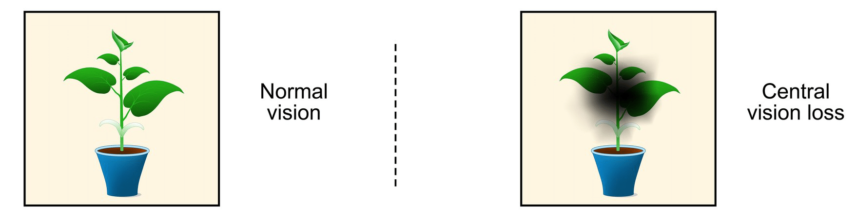
Left: normal vision. Right: what central vision loss from macular degeneration can look like. Side vision stays intact.
While most vision loss from macular degeneration is gradual, sometimes there can be an abrupt change. In some cases, the first symptoms of macular degeneration will be the sudden onset of distortion or a blind spot in the vision.
Checking Your Vision at Home
An Amsler grid is a simple square of graph paper with a dot in the center. It is the easiest way to catch a change in your central vision between visits. Hold the grid 12 to 15 inches from your face in good light, cover one eye, and look at the center dot. Then repeat with the other eye. Every line should look straight and every square should be the same size. If any lines look wavy, dark, or missing, call us the same day.

Cover one eye, hold the grid at reading distance, and look at the center dot.
Download a printable Amsler grid here, or use the one the American Academy of Ophthalmology provides with instructions. If you have been diagnosed with macular degeneration, check each eye once a day. It takes less than a minute.
Which Type of Macular Degeneration Do I Have?
Age-related macular degeneration comes in two flavors, dry and wet. While most folks think of dry macular degeneration as ‘the good kind’ and wet macular degeneration as ‘the bad kind,’ you can read below that this distinction is not entirely helpful.
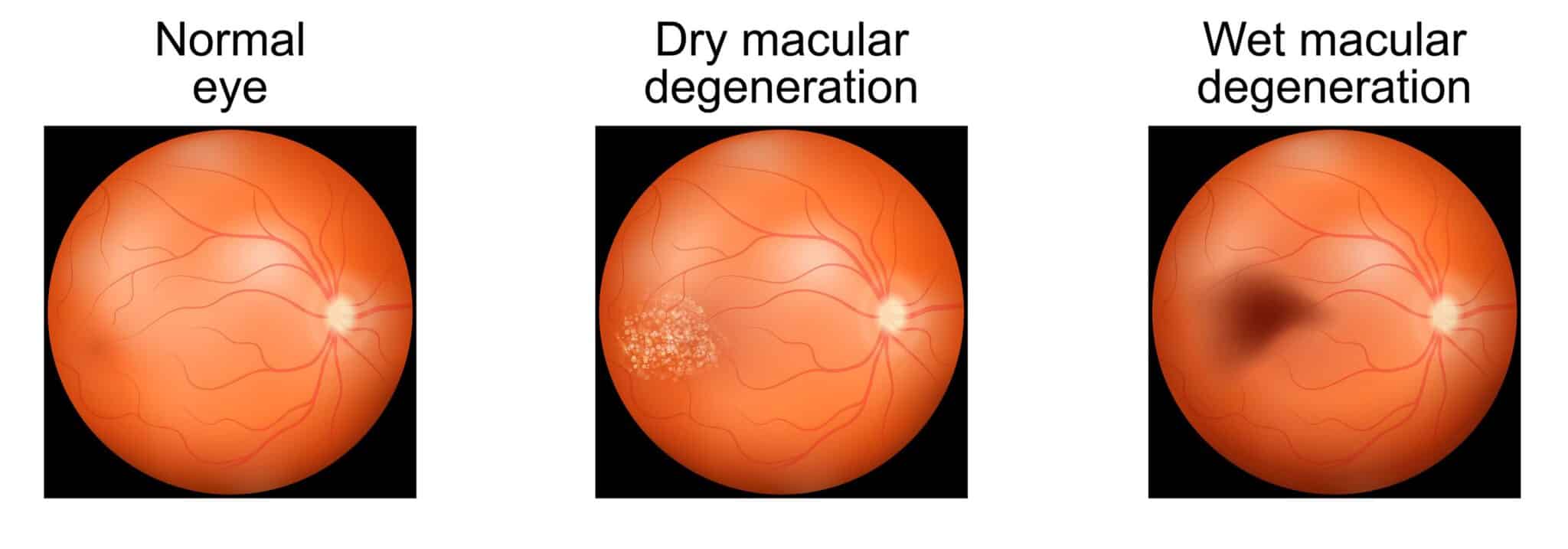
Left to right: a normal retina, dry macular degeneration with drusen deposits, wet macular degeneration with bleeding under the retina.
Dry Macular Degeneration
Dry macular degeneration is the most common form of the disease, affecting 90% of patients with A.M.D. Dry macular degeneration is characterized by the presence of bumps underneath the retina, called drusen. These retinal changes show up early in the disease and typically take a long time to lead to vision loss. The size, number, and appearance of drusen can be prognostic for future vision loss. As the years pass by, the drusen typically become more numerous and destructive to the overlying retina. Eventually, the bumps may disappear leaving blind spots in their place.
Wet Macular Degeneration
Wet macular degeneration is less common than dry, affecting only 10% of patients. Wet macular degeneration is characterized by the growth of new blood vessels that leak and bleed under the retina. When left untreated, these changes lead to rapid retinal damage and vision loss.
Why “Dry” Does Not Mean “Safe”
Although wet macular degeneration can lead to rapid vision loss, it is entirely treatable. Patients who get timely treatment for wet macular degeneration can maintain their vision for years until their underlying dry macular degeneration takes over (yes, you can have wet and dry macular degeneration at the same time).
Treatments for dry macular degeneration are more limited. Vitamins can lower the risk of progression for some patients, and newer injections can slow the growth of the blind spots of advanced dry disease, but nothing yet restores the vision that dry macular degeneration takes away. It’s for these reasons that just as many patients lose vision from dry macular degeneration as they do wet.
The Stages of Macular Degeneration
Retina specialists describe macular degeneration in three stages. Knowing your stage explains why one patient is told to take vitamins and watch, while another needs treatment right away.
- Early: A few medium-sized drusen are present. Most people have no symptoms and find out only through a dilated eye exam.
- Intermediate: Drusen are larger or more numerous, or the pigment layer under the retina has started to change. Some people notice mild blurring or trouble seeing in low light. Others still notice nothing. This is the stage where AREDS2 vitamins can lower the risk of progression.
- Advanced: Vision loss has begun. Advanced disease takes one of two forms: geographic atrophy, in which patches of the retina have worn away (advanced dry A.M.D.), or wet A.M.D., in which abnormal blood vessels have started to leak.
Any stage of dry macular degeneration can turn into wet macular degeneration. That is why regular monitoring matters even when your vision feels stable.
What Causes Age-Related Macular Degeneration?
There is no ‘one’ cause for macular degeneration, just a list of risk factors that increase your risk for developing the condition. Besides age >60, the most common risk factors for age-related macular degeneration include family history and smoking. Other potential contributing factors include:
- Female gender
- Caucasian race
- Diabetes
- Obesity
- High blood pressure
- Excessive sun exposure
- Poor nutrition
How is Macular Degeneration Diagnosed?
Regular eye examinations become more important as we get older, so that age-related vision changes, like A.M.D., can be detected and treated as early as possible. Macular degeneration can be diagnosed by an ophthalmologist during a dilated eye exam with the help of retinal imaging called optical coherence tomography (OCT).
What to Expect at Your Visit
Your visit begins with a vision check and drops to dilate your pupils. Dilation lets your retina specialist see the macula clearly and takes about 20 to 30 minutes to work. Your near vision will stay blurry and light-sensitive for four to six hours afterward, so bring sunglasses and a driver if you can.
An OCT scan takes a cross-section picture of the macula in a few seconds, without touching the eye. It shows drusen, thinning of the retina, and any fluid or bleeding from wet macular degeneration. If wet disease is suspected, your doctor may also order fluorescein angiography, a series of photographs taken after a dye is injected into a vein in your arm, to map the leaking vessels.
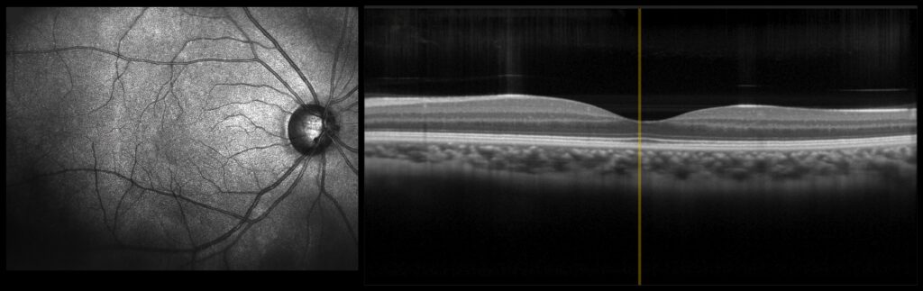
An OCT scan of a healthy macula. The dip in the center is the fovea. In macular degeneration, this scan shows drusen, thinning, or fluid under the retina.
How often you are seen depends on your stage. Patients with early or intermediate disease are usually checked every six to twelve months. Patients being treated for wet macular degeneration are seen more often, on a schedule set by their retina specialist.
How is Macular Degeneration Treated?
Treatment depends on which type you have and how far it has progressed. There is no cure yet, but modern treatment can slow the disease and, in wet macular degeneration, often stabilize or improve vision. Our macular degeneration treatment page covers each option in detail. Here is the short version.
Treating Wet Macular Degeneration
Wet macular degeneration is treated with eye injections of anti-VEGF medication. VEGF, short for vascular endothelial growth factor, is the signal that tells abnormal blood vessels to grow and leak. Blocking it stops the leaking and lets the retina dry out. The medications used include Eylea, Lucentis, Avastin, and Vabysmo. The injections wear off, so treatment is repeated on a schedule, often every one to four months. For some patients, the Susvimo implant can release medication continuously and reduce the number of visits.
Treating Dry Macular Degeneration
For intermediate dry macular degeneration, the AREDS2 vitamin formula lowers the risk of progressing to advanced disease by about 25% in appropriate patients. For advanced dry macular degeneration with geographic atrophy, two injectable medications, Syfovre and Izervay, can slow the growth of the damaged areas. Some patients with dry disease are also candidates for Valeda light therapy, a painless in-office treatment that uses specific wavelengths of light to support the cells of the macula.
Living Well with Lower Vision
When central vision has been lost, low-vision tools such as brighter lighting, magnifiers, and specialized glasses can make reading and daily tasks easier. A low-vision specialist, usually an optometrist with additional training, can match these tools to your needs.
Will I Go Blind from Macular Degeneration?
Almost never. Macular degeneration damages central vision, but it leaves side vision intact. Even patients with advanced disease can usually walk through a room, see shapes and movement, and take care of themselves. What they lose is the fine detail needed for reading, driving, and recognizing faces.
The bigger risk is delay. Wet macular degeneration caught early and treated consistently can hold reading and driving vision for years. Wet macular degeneration left alone for months can cause permanent scarring. If you notice a change, do not wait to see whether it passes.
For Family Members and Caregivers
Often it is an adult son or daughter who notices the first signs. Watch for a parent who holds reading material closer, needs a brighter lamp, misses words on a page, stops recognizing faces across a room, or quietly gives up night driving. Any of these is a reason to schedule a retinal exam.
Once macular degeneration is diagnosed, the most helpful things you can do are practical. Help with the daily Amsler grid check. Keep a current list of eye drops and medications. Plan a ride home from dilated visits and injection appointments. Family members are welcome at appointments, and our doctors are glad to explain the findings to both of you.
When to Call a Retina Specialist:
Most macular degeneration changes slowly. A sudden change is different, and it is often the first sign that dry macular degeneration has turned wet. Wet macular degeneration responds best when treatment starts quickly.
Call us right away if you notice:
- Straight lines that suddenly look wavy, bent, or broken
- A new dark, gray, or blank spot in the center of your vision
- A sudden drop in vision in one eye, even if the other eye seems fine
- Words or letters that seem to disappear while you read
- Any new change on your Amsler grid
If you experience any of these symptoms, do not wait for your next scheduled appointment. Call our office at (941) 351-1200. We reserve time each day for urgent retinal evaluations, and our team will work to get you seen as quickly as possible.
While most vision loss from macular degeneration is gradual, sometimes there can be an abrupt change. In some cases, the first symptoms of macular degeneration will be the sudden onset of distortion or a blind spot in the vision.
Macular Degeneration Specialist and founder of Shane Retina
Macular Degeneration FAQs
What is macular degeneration?
Macular degeneration, also called age-related macular degeneration (AMD), is an eye disease that damages the macula, the part of the retina responsible for sharp central vision. It causes gradual loss of central vision, making reading, driving, and recognizing faces more difficult.
The macula sits at the center of the retina and allows you to see fine detail and color clearly. In macular degeneration, the cells of the macula gradually deteriorate over time. The disease most commonly affects people over the age of 60 and becomes more prevalent with advancing age.
There are two main forms of macular degeneration: dry and wet. Dry macular degeneration progresses slowly and is more common, while wet macular degeneration involves abnormal blood vessel growth and can cause more rapid vision changes. Although peripheral vision is usually preserved, untreated central vision loss can significantly limit daily activities.
Early diagnosis and ongoing follow-up help protect remaining central vision.
What are the first signs of macular degeneration?
The first signs of macular degeneration are usually subtle changes in your central vision. Many patients first notice that straight lines appear slightly wavy, words seem distorted, or they need brighter light to read comfortably.
Other early symptoms may include:
- Blurry or fuzzy central vision
- Difficulty recognizing faces
- Trouble adapting to low light
- A small dim or gray spot in the center of vision
In dry macular degeneration, these changes typically develop gradually over months or years. In wet macular degeneration, vision can change more suddenly, sometimes over days to weeks.
Because early macular degeneration may cause only mild or intermittent symptoms, regular dilated eye examinations by an ophthalmologist are important for early detection. Identifying changes early gives you the best chance of maintaining useful central vision.
What does vision look like with macular degeneration?
With macular degeneration, central vision often becomes blurred, distorted, or partly missing. Straight lines may look wavy, faces may appear unclear, and a dark or gray spot can develop in the middle of your visual field.
In early stages, patients often notice mild distortion when reading or looking at grid patterns. Letters may seem bent or parts of words may appear faded. As the condition progresses, the central blind spot can enlarge, making detailed tasks like reading, driving, or recognizing faces increasingly difficult.
Peripheral, or side vision, usually remains intact. Even in advanced stages, most patients can still navigate a room and maintain orientation.
However, loss of central clarity can significantly affect daily independence if not addressed.
Prompt evaluation of new distortion or sudden central vision changes is important, especially because wet macular degeneration can progress more quickly than the dry form.
What causes macular degeneration?
Macular degeneration is caused by a combination of aging, genetic factors, and environmental influences that gradually damage the cells of the macula. There is no single cause, but several risk factors increase the likelihood of developing the condition.
The strongest risk factor is age. As we get older, the light-sensitive cells in the macula become more vulnerable to wear and tear. Over time, metabolic waste can accumulate beneath the retina in the form of drusen, small yellow deposits associated with dry macular degeneration. In some individuals, abnormal blood vessels grow beneath the retina, leading to wet macular degeneration.
Other major risk factors include:
- Family history of macular degeneration
- Smoking
- High blood pressure
- Elevated cholesterol
- Obesity
- Poor diet
- Excessive sun exposure
Genetics also plays an important role. Certain inherited variations in immune system pathways have been linked to higher risk for advanced disease.
While aging cannot be prevented, managing cardiovascular risk factors, avoiding smoking, and keeping up with routine eye exams may help reduce the risk of severe vision loss from AMD.
Is macular degeneration hereditary?
Yes, macular degeneration can run in families. Having a close relative with age-related macular degeneration increases your risk of developing the condition, especially in its more advanced stages.
Research has shown that certain genetic variations, particularly in parts of the immune system involved in inflammation and complement pathways, are associated with a higher risk of macular degeneration. However, genetics is only part of the story. Not everyone with a family history will develop the disease, and lifestyle factors such as smoking, diet, and cardiovascular health also play an important role.
If you have a parent or sibling with macular degeneration, it is especially important to have regular dilated eye examinations. Early detection allows changes to be monitored closely and treated at the right time.
What is the difference between dry and wet macular degeneration?
Dry and wet macular degeneration affect the same area of the retina, but wet AMD can cause faster vision changes and usually requires prompt treatment. Dry macular degeneration is more common and progresses slowly. Wet macular degeneration is less common but can cause more rapid vision changes.
Dry macular degeneration accounts for about 85 to 90 percent of cases. It is characterized by the buildup of small yellow deposits under the retina called drusen and gradual thinning of the macular cells. Vision loss typically develops slowly over years.
Wet macular degeneration occurs when abnormal blood vessels grow beneath the retina and begin to leak fluid or blood. This leakage can cause sudden distortion, blurry vision, or a dark spot in the center of vision. Although wet macular degeneration can progress quickly, it is treatable with modern injection therapies that can stabilize vision and improve it in many cases. Treatment is usually ongoing, and results vary based on how advanced the disease is at diagnosis.
Some patients with dry macular degeneration can later develop the wet form. Because of this, regular monitoring is essential so that treatment can begin promptly if changes occur.
How fast does macular degeneration progress?
Macular degeneration usually progresses slowly over many years, especially in the dry form of the disease. Most patients experience gradual changes in central vision rather than sudden, complete vision loss.
In early and intermediate dry macular degeneration, vision may remain stable for long periods. Changes often develop subtly, such as needing brighter light to read or noticing mild distortion. Progression from early to advanced stages can take several years or even decades.
Wet macular degeneration behaves differently. When abnormal blood vessels develop under the retina, vision changes can occur more quickly, sometimes over days or weeks. Prompt treatment with injection therapy can often stabilize or improve vision if started early.
The rate of progression varies widely between individuals and depends on factors such as genetics, smoking history, cardiovascular health, and how closely the condition is monitored. Routine retina examinations allow changes to be detected early so treatment can begin at the right time.
Can macular degeneration be reversed or cured?
Macular degeneration cannot currently be cured, and existing damage to the retina cannot be fully reversed. However, modern treatments can slow progression, stabilize vision, and in some cases improve visual clarity, especially in wet macular degeneration.
In dry macular degeneration, vitamin therapy such as the AREDS2 formula can reduce the risk of progression in intermediate stages. Newer therapies, including complement inhibitors (Syfovre and Izervay) for advanced dry AMD, can slow the growth of blind spots. While these treatments do not restore lost cells, they can help preserve remaining vision for longer.
In select patients with earlier-stage disease, light-based treatments such as Valeda photobiomodulation therapy may also be used to support retinal function and slow progression.
Wet macular degeneration is treatable with anti-VEGF eye injections. These medications can stop leakage from abnormal blood vessels and often improve vision when started early. Many patients maintain reading and driving vision for years with consistent treatment.
Although macular degeneration cannot be cured, early diagnosis and consistent care can significantly improve long-term visual outcomes. Regular monitoring allows treatment to begin at the optimal time to protect central vision.
What is the best eye vitamin for macular degeneration?
The most studied eye vitamin for macular degeneration is the AREDS2 formula, which can lower the risk of progression in appropriate patients with intermediate AMD.
AREDS2 contains vitamin C, vitamin E, zinc, copper, lutein, and zeaxanthin. These nutrients help reduce oxidative stress in the retina and support the health of macular cells. Studies from the Age-Related Eye Disease Study demonstrated that this formula can lower the risk of progressing to advanced disease by about 25 percent in appropriate patients.
It is important to understand that AREDS2 does not prevent macular degeneration and does not restore lost vision. It is most beneficial for patients with intermediate dry AMD or advanced disease in one eye. It is not typically recommended for early-stage AMD without retinal changes.
Before starting any supplement, it is best to confirm your stage of macular degeneration with a retina specialist to ensure that AREDS2 is appropriate for you.
What happens if you stop eye injections for macular degeneration?
If you stop receiving eye injections for wet macular degeneration, abnormal blood vessels can begin leaking again, leading to renewed swelling, bleeding, and potential permanent vision loss. These injections control the disease, but they do not cure it.
Anti-VEGF medications work by blocking the signals that cause harmful blood vessels to grow under the retina. When treatment is paused for too long, those signals can become active again. Over weeks to months, fluid may reaccumulate in the macula, and central vision can decline.
In some cases, vision loss that occurs after stopping injections may not fully recover, even if treatment is restarted. That is why staying on the recommended treatment schedule is critical for maintaining reading and driving vision.
If scheduling, transportation, or injection frequency is becoming difficult, speak with your retina specialist. There may be extended-interval treatments or long-acting options available that can reduce treatment burden while maintaining visual stability.
Does macular degeneration cause blindness?
Macular degeneration can cause significant central vision loss, but it rarely causes complete blindness. The disease primarily affects the macula, which controls detailed, straight-ahead vision, while peripheral vision usually remains intact.
In advanced stages, especially if untreated, patients may develop a large blind spot in the center of their vision. This can make reading, driving, and recognizing faces very difficult.
However, side vision is typically preserved, allowing individuals to navigate their surroundings independently.
The risk of severe central vision loss has decreased over time due to modern treatments. Anti-VEGF injections for wet macular degeneration and newer therapies for advanced dry AMD have significantly improved long-term outcomes.
Early diagnosis and timely treatment are the most important factors in reducing the risk of severe vision impairment. With proper care, many patients maintain functional vision for years after diagnosis.
Reviews of Our Macular Degeneration Services
Patients with macular degeneration see us often, sometimes every month for years, so the relationship matters. Our patients consistently describe Dr. Shane and our team as thorough, patient, and clear about what is happening and why. Many have been with us since their diagnosis and have brought spouses, friends, and neighbors along the way.
The reviews below are a small sample of what real patients have said about their macular degeneration care at Shane Retina.
Your Shane Retina Care Team
Macular degeneration is diagnosed and managed at Shane Retina by our team of fellowship-trained retina specialists. Each of our physicians brings the clinical experience that complex retinal conditions require.
Thomas Shane, M.D.
Anita Shane, M.D.
Konica Singla, M.D.
Maria Castaños, M.D.
Gabriel Dib, FNP-C
Ayah Etyem, PA-C
Where to Get Macular Degeneration Care in Sarasota and Venice
You can receive expert macular degeneration evaluation and treatment at any of our Shane Retina locations in throughout Sarasota and Venice, Florida. Each office offers comprehensive retina care, advanced diagnostic imaging, and proven treatment options in a calm and efficient clinical setting.
Sarasota - University Parkway Office
Located just west of I-75 on University Parkway in University Health Park.
Our University Parkway location serves patients from Sarasota, Lakewood Ranch, Bradenton, and nearby communities. This office provides comprehensive evaluation and treatment for both dry and wet macular degeneration.
Sarasota - South Tamiami Trail Office
Located on South Tamiami Trail just south of Bee Ridge Road.
Our South Tamiami Trail location serves patients throughout Sarasota, Siesta Key, and surrounding neighborhoods. This office offers full evaluation and treatment for macular degeneration, with a focus on ongoing care and clear communication.
Venice Retina Office
Located in central Venice near Venetia Bay Boulevard.
Our Venice Retina location serves patients in Venice, North Port, Englewood, and nearby communities. This office provides comprehensive evaluation and treatment for macular degeneration, including ongoing monitoring and advanced therapies.
Schedule Your Macular Degeneration Evaluation Today
For those lucky enough to live in Sarasota, Manatee County, or Venice, the renowned staff at Shane Retina is available to help with your macular degeneration evaluation and treatment. Our retina specialists, led by Dr. Thomas Shane M.D., will make sure you get the right treatment you need.
If you are experiencing any A.M.D. symptoms, or have a concern about your eye health, contact us today to set up an appointment at our Sarasota or Venice offices. Dr. Shane works hard to be available to our patients to answer questions and calm concerns.
Call us at (941) 351-1200 or schedule online.