Retinal Artery Occlusion Diagnosis and Emergency Care in Sarasota, FL

Sudden vision loss in one eye should be evaluated immediately. Early action can help protect your sight and overall health.

A retinal artery occlusion, often called an “eye stroke,” occurs when blood flow to the retina is suddenly blocked. This causes rapid, painless vision loss and may signal a higher risk for stroke in the brain.

Our ophthalmologists diagnose this condition urgently using advanced imaging and work closely with local specialists to ensure you receive the medical attention you need right away.

Retinal artery occlusion can be a life-threatening condition that causes severe and permanent loss of vision. Patients who experience an ‘eye stroke’ are at high risk for a stroke in the brain. The ophthalmologists at Shane Retina are specialized in diagnosing and treating retinal artery occlusion. If you believe that you are experiencing an eye stroke in Sarasota or Manatee County, please call us for an appointment before reading the rest of this section.

Understanding Retinal Artery Occlusion

To understand retinal artery occlusion, it is important to know the anatomy of blood flow to the eye. Beginning in the heart, blood is pumped through the aorta to the right and left carotid arteries on the way to the eye. The eyeball is fed by the ophthalmic artery, with most of the retinal blood flow coming from a single branch called the central retinal artery. The remainder of the blood flowing through the carotid artery continues to the brain.

The central retinal artery enters through the optic nerve in the back of the eye. After it reaches the retina, it branches into an upper and lower section that wraps around the center of the vision. From there, the branch retina arteries further divide into smaller and smaller capillaries.

What is An Eye Stroke?

Retinal Artery Occlusion Spcialist in Sarasota Florida Unlike in the brain, the blood flow to the retina is one-way. There are no redundant connections between branches of the artery. If a retinal artery becomes blocked, all the blood flow downstream is stopped. When blood flow stops to the retina, permanent damage and loss of vision occur over the span of an hour. This is typically referred to as an eye stroke or retinal artery occlusion.

There are many possible causes for a blockage of the retinal artery. The most common cause involves a particle of cholesterol that breaks off from the carotid artery wall and travels into the central retinal artery. At some point, the particle called an embolus, lodges in the vessel, and plugs the blood flow.

In other cases of eye stroke, a clot forms in the heart or carotid artery and travels to the eye. This collection of platelets and protein similarly can occlude the retinal vessels. Rarely, clots or particles in the veins can cross over into the arterial system through a defect in the heart. This is particularly likely when patients inject contaminated street drugs into their veins.

Some patients experience an artery occlusion without having an embolus journey from the heart or carotid artery. Instead, the retinal artery becomes inflamed due to an autoimmune reaction. This is called temporal arteritis or giant cell arteritis. When the vessel becomes inflamed, the inside of the vessel narrows until no blood flow can pass through.

What Are the Risk Factors for Retinal Artery Occlusion?

Anything that causes the buildup of cholesterol in your arteries increases your risk for retinal artery occlusion. This includes conditions such as high blood pressure, elevated cholesterol, diabetes, smoking, obesity, poor diet, and lack of exercise. When cholesterol accumulates in the walls of your carotid arteries, it is more likely to break free and lodge in a retinal artery downstream.

Anything that increases your risk for arterial clot formation also can lead to retinal artery occlusion. This includes patients who undergo cardiac valve or carotid artery procedures. Clotting disorders, trauma, and atrial fibrillation sometimes lead to loss of vision through the formation of clots that lodge in the retinal arteries.

For giant cell arteritis, advanced age is the main risk factor. Those patients with polymyalgia rheumatica are at particularly high risk for inflammatory blockage of a retinal artery.

What Are the Symptoms of a Retinal Artery Occlusion?

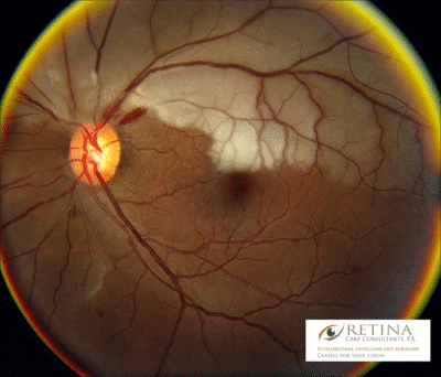
Retinal artery occlusion causes sudden, painless loss of vision in one eye. Patients will complain of a graying or blacking out of vision. In the case of central retinal artery occlusion, the entire visual field is lost. With a branch retinal vein occlusion, only a portion of the vision is lost, usually above or below the horizontal midline. Sometimes, the blockage of blood flow is only partial, and patients will experience a recurrent, temporary loss of vision with recovery minutes later.

In cases of retinal artery inflammation, the patient may experience headaches, jaw pain, or tenderness over the temples along with vision loss. Unlike with an embolus, temporal arteritis may cause loss of vision in both eyes simultaneously.

How is a Retinal Artery Occlusion Diagnosed?

Retinal Artery Occlusion Spcialist in Sarasota Florida Anyone suspected of having a retinal artery occlusion should seek a same-day dilated examination with an ophthalmologist. During the exam, your vision will be measured, pupils dilated, and retinal images are taken. The eye doctor will examine your retinas and determine whether a blockage of blood flow has occurred. In most cases, the retina will appear pale where the loss of blood flow has caused swelling of the nerve fibers. Sometimes, the ophthalmologist can spot a particle of cholesterol within the retinal artery. 

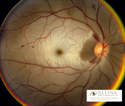
Sometimes a specialized imaging technique called fluorescein angiogram is helpful. During this test, dye is injected into an arm vein, and photos are taken as the dye passes through the retinal circulation. If an eye stroke has occurred, there will be a portion of the retina that does not light up with dye downstream of the blockage.

When retinal artery inflammation is suspected (temporal arteritis or giant cell arteritis), urgent lab testing is obtained. These tests (ESR and CRP) measure inflammation in the bloodstream. If these lab tests are elevated, the patient will be sent for a biopsy of the temporal artery for a definitive diagnosis of arterial inflammation.

Shane Retina, has ophthalmologists specializing in managing central retinal artery occlusions. If you experience a sudden loss of vision, seek an immediate dilated eye examination with us at one of our Sarasota and Manatee Count office locations. This exam may involve dilation, blood tests, photographs, or other specialized imaging tests of the retina.

Get Immediate Help for Your Eye Stroke

Retinal artery occlusion may be a warning sign for a life-threatening condition, so do not delay immediate medical attention if you are experiencing the symptoms mentioned above. Contact us for retinal artery occlusion treatment today in Sarasota, Bradenton, and Lakewood Ranch, FL. Come to one of our two convenient locations in the area and we will give you the immediate care you need for your eye stroke.

How is a Retina Artery Occlusion Treated?

A new retinal artery occlusion indicates that a patient is at high risk for an acute stroke in the brain.  In fact, up to 5% of patients will experience a brain stroke within a couple of weeks of their eye stroke.  Therefore, anyone who is diagnosed with a new retinal artery occlusion is typically sent to the emergency room for stroke evaluation.  While at the emergency department, ultrasound is used to image the carotid arteries and heart to make sure there are not any significant risks for additional stokes.

Many treatments have been attempted to dislodge retinal artery occlusions before permanent damage has been done.  These include massaging the eye, breathing carbon dioxide, removing eye fluid with a needle, injecting clot-busting medication, and surgically removing the clot.  Unfortunately, no intervention has been shown to reliably restore vision when compared to observation.   Furthermore, most interventions would need to occur within one hour of the loss of vision to be effective.

In cases of suspected giant cell arteritis, patients are treated with oral steroids while they await the results of the temporal artery biopsy.  If the biopsy is positive, the steroids are continued for many months to avoid the risk of sudden, severe vision loss in both eyes.  Recently, an alternative to steroid medication was FDA approved for use in temporal arteritis.  This new medicine may help to avoid the many side effects of steroid medication.

Will I Regain My Sight After Experiencing a Retinal Artery Occlusion?

Unfortunately, vision loss from retinal artery occlusion is severe and persistent.  While patients will typically experience improvement of the vision over time, much of the sight does not return.  Still, it is important for these patients to be examined with dilation from time to time to ensure that additional blockages or blood vessel growth do not occur.

Our Retinal Artery Occlusion Treatment

Retinal Artery Occlusion Spcialist in Sarasota Florida Shane Retina, has ophthalmologists specializing in managing central retinal artery occlusions. If you experience a sudden loss of vision, seek an immediate dilated eye examination with us at one of our Sarasota and Manatee Count office locations. This exam may involve dilation, blood tests, photographs, or other specialized imaging tests of the retina.


Retinal Artery Occlusion FAQs

Click on a question below to view the answer.

Retinal artery occlusion is a sudden blockage of blood flow to the retina, often referred to as an “eye stroke.” When blood supply is interrupted, the retinal tissue is deprived of oxygen, leading to rapid and often severe vision loss in one eye.

The retina depends on continuous blood flow to function properly. If the central retinal artery or one of its branches becomes blocked by a clot or cholesterol plaque, the affected portion of the retina can suffer permanent damage quickly, sometimes within hours.

Retinal artery occlusion is a medical emergency not only because of the risk to vision, but also because it may signal an increased risk of stroke elsewhere in the body. Immediate evaluation is critical to protect both vision and overall health.

Yes, central retinal artery occlusion is considered a type of stroke that affects the eye. It occurs when blood flow through the main artery supplying the retina is suddenly blocked, similar to how a clot can block blood flow to the brain.

Because the retina is part of the central nervous system, this event is managed similarly to a stroke affecting the brain. In addition, patients who experience a retinal artery occlusion are at increased risk of having a stroke in the brain, especially within the first few days to weeks after vision loss.

For this reason, central retinal artery occlusion is treated as a medical emergency. Patients are often referred immediately for stroke evaluation, including imaging of the carotid arteries and heart, to identify the source of the blockage and reduce the risk of further events.

Sudden, painless loss of vision in one eye should never be ignored, as it may represent both an eye emergency and a warning sign of a more serious vascular condition.

Retinal artery occlusion is most commonly caused by a blood clot or cholesterol plaque that travels to and blocks one of the arteries supplying the retina. This blockage deprives the retinal tissue of oxygen and leads to sudden vision loss.

The most frequent source of the blockage is a cholesterol embolus that breaks loose from the carotid arteries in the neck. Clots can also originate from the heart, particularly in patients with atrial fibrillation or other cardiac conditions. Less commonly, inflammation of the arteries, known as giant cell arteritis, can narrow or close off the retinal artery.

Risk factors for retinal artery occlusion are similar to those for stroke and heart disease. They include high blood pressure, high cholesterol, diabetes, smoking, obesity, and cardiovascular disease. Because the condition often signals an underlying vascular problem, patients diagnosed with retinal artery occlusion typically undergo urgent medical evaluation with a retina specialist to reduce the risk of stroke.

No, retinal artery occlusion is typically not painful. It usually causes sudden, painless loss of vision in one eye.

Patients often describe their vision as going gray, dim, or completely dark without any warning or discomfort. Because there is no pain, some people delay seeking care, which can be dangerous.

An important exception is when retinal artery occlusion is caused by giant cell arteritis, an inflammatory condition affecting the arteries. In those cases, patients may experience symptoms such as headache, scalp tenderness, jaw pain while chewing, or general fatigue along with vision loss. This form requires immediate medical treatment to prevent vision loss in the other eye.

Any sudden loss of vision, even without pain, should be treated as a medical emergency and evaluated immediately.

Retinal artery occlusion is diagnosed through an urgent dilated eye examination combined with retinal imaging. Because the condition is a medical emergency, evaluation should occur the same day sudden vision loss develops.

During the exam, the ophthalmologist measures vision, dilates the pupils, and carefully examines the retina. In many cases, the retina appears pale due to lack of blood flow, and a small cholesterol plaque may sometimes be visible inside the blocked artery.

Additional imaging, such as fluorescein angiography, may be performed to assess blood flow through the retinal vessels. If inflammation such as giant cell arteritis is suspected, blood tests are ordered immediately to measure markers of inflammation, and further testing may follow.

Because retinal artery occlusion can signal an increased risk of stroke, patients are often referred for urgent medical evaluation, including imaging of the carotid arteries and heart. Prompt diagnosis is critical not only for vision but also for overall health.

Retinal artery occlusion cannot be cured once permanent damage to the retina has occurred. When blood flow is blocked for more than a short period of time, the retinal tissue suffers irreversible injury.

Because the retina depends on a constant supply of oxygen, vision loss can become permanent within hours. While some patients may experience slight improvement over time, most do not regain the vision that was lost at the time of the event.

Although the vision itself often cannot be restored, the underlying cause should be treated. Identifying and managing cardiovascular risk factors such as high blood pressure, cholesterol, diabetes, or heart rhythm abnormalities is critical to reducing the risk of stroke or additional vascular events.

In cases caused by inflammatory conditions such as giant cell arteritis, prompt treatment with steroids can prevent vision loss in the other eye.

While retinal artery occlusion is not curable, urgent medical evaluation can protect overall health and reduce the risk of future, potentially life-threatening complications.

Get Immediate Help for Your Eye Stroke

Retinal artery occlusion may be a warning sign for a life-threatening condition, so do not delay immediate medical attention if you are experiencing the symptoms mentioned above. Contact us for retinal artery occlusion treatment today in Sarasota, Bradenton, and Lakewood Ranch, FL. Come to one of our two convenient locations in the area and we will give you the immediate care you need for your eye stroke.