
Retinal Vein Occlusion Diagnosis and Treatment in Sarasota, FL
Early evaluation and proper treatment can help protect your central vision.
A retinal vein occlusion occurs when a vein that drains blood from the retina becomes blocked, leading to sudden, painless blurring of vision in one eye. This can cause swelling and bleeding in the macula, making reading or detailed vision more difficult. Our retina specialists use advanced imaging to determine the type of vein occlusion and recommend treatments that help stabilize and improve vision.
If you notice a sudden change in vision, especially in one eye, a prompt dilated exam is important. Early diagnosis gives you the best chance of maintaining the clearest vision possible.
What is a Retinal Vein Occlusion?
A retinal vein occlusion is the sudden blockage of blood flow out of the eyeball, which causes macular bleeding, swelling, and loss of vision.
The retina is the part of the eye that receives light and transmits images to the brain. The eye requires a lot of energy to create vision, so there is a significant amount of blood flow bringing oxygen and nutrients to and from the retina. Two major vessels, called the central retinal artery and vein enter the eye through the optic nerve and branch over the surface of the retina. These vessels are the only source of blood flow for most of the inner retina.
As our retinal blood vessels age, they accumulate cholesterol causing hardening of artery walls. Our blood may also develop an increasing propensity to clot. Sometimes these factors combine in the eye to cause a sudden blockage of a retinal vein, which prevents blood flow from leaving the retina. The blockage, or ‘occlusion,’ results in reduced retinal blood flow, retinal bleeding, swelling, and sudden painless loss of vision.
Compare Central Retinal Vein Occlusion and a Branch Retinal Vein Occlusion
Retinal vein occlusion can be subdivided into two categories, depending at what point a clot forms along the network of blood vessels in the eye. A central retinal vein occlusion (CRVO) occurs when the single, large vein that drains blood from the inner retina becomes blocked. This usually occurs as the vein passes through the optic nerve on its pathway out of the eye. Because this vein is the only pathway for blood to leave the eye, hemorrhages form throughout the retina, blood flow is significantly reduced, and swelling throughout the center of the retina (macula) occurs.
Central retinal vein occlusions can be classified based on how much they restrict blood flow to the eye. Mild CRVO’s cause only minor disruption of vision and typically require fewer treatments to improve. Severe CRVO’s lead to profound loss of blood flow to the eye, which in turn requires intensive treatment to prevent severe vision loss.
In contrast to a CRVO, a branch retinal vein occlusion (BRVO) occurs when only a tributary of the main retinal vein is obstructed. This typically occurs at a point where a branch of the retinal artery and retinal vein cross each other along the surface of the retina. At this crossing point, the stiff-walled artery can press on the soft-walled vein, causing the vein to collapse and obstruct the blood flow through the vessel. The resulting hemorrhages and swelling within a section of the retina can harm vision, but do not affect the entire visual field like a CRVO.
What are the Risk Factors for Retinal Vein Occlusion?
The risk factors for a retinal vein occlusion include anything that increases the stiffness of artery walls or elevates the propensity for forming blood clots. Factors that lead to hardening of the arteries include all of the familiar cardiovascular risk factors for heart attack and stroke: Blood pressure, cholesterol, diabetes, smoking, diet, exercise, weight, and family history. In younger patients, the propensity to form blood clots can be elevated by risk factors such as smoking, estrogen use, inherited clotting disorders, autoimmune conditions, and cancer.
In patients over the age of 50, the most common causes for retinal vein occlusion are cardiovascular risk factors. These folks should be sent for a primary care doctor evaluation of their blood pressure, cholesterol, blood sugar, and counseling regarding their cardiovascular risk profile. Anyone who experiences a retinal vein occlusion under the age of 50 should also be evaluated for clotting disorders, autoimmune conditions, and blood disorders including cancer.
What are the Symptoms of a Retinal Vein Occlusion?
A retinal vein occlusion causes a sudden, painless blurring of vision in one eye. While a central retinal vein occlusion blurs all of the central vision, a branch retinal vein occlusion may only affect the upper visual field or lower visual field. The blurriness from retinal vein occlusion will not fluctuate, but rather persist for months or years if not treated promptly.
How is a Retinal Vein Occlusion Diagnosed?
If you suspect that you may have a retinal vein occlusion, seek a dilated examination with and ophthalmologist specialized in macula and retina conditions. During your visit, you will undergo specialized imaging that can evaluate whether retinal swelling is causing vision loss. The ophthalmologist will examine your retina and discuss treatment options with you.
If you are under the age of fifty or suspected to have an underlying systemic cause for your retinal vein occlusion, you may be sent for further laboratory testing or imaging. All patients who experience a retinal vein occlusion should have regular follow-up with their primary care doctor to monitor and treat cardiovascular risk factors.
How is Retina Vein Occlusion Treated?
A retinal vein occlusion is most commonly treated with injections of medicine into the eye. The medicine is specifically targeted to treat swelling of the retina, which can restore and protect central vision. While the injections have to be repeated every 4-12 weeks over months or years, they have been proven to prevent severe vision loss that can occur without treatment.
Sometimes, a retinal vein occlusion may be treated with laser. Typically, the laser is used in cases where injections are ineffective (rare) or when there is new blood vessel growth in the retina that can cause bleeding, glaucoma, or retinal detachment. While there are no surgeries to remove clots from a retinal vein, occasionally the surgical removal of blood from the eye is warranted.
Lastly, retinal vein occlusions that occur without symptoms, happen outside of the center of vision, or don’t cause retinal swelling can be safely monitored without treatment.
What is the Prognosis for a Retinal Vein Occlusion?
With appropriate and timely treatment, excellent vision can be achieved even after a retinal vein occlusion. Conversely, the most common cause for poor vision after a retinal vein occlusion is delay in diagnosis or treatment. Even in some cases of excellent follow-up however, the retinal vein occlusion is unresponsive to medical treatment due to severely reduced blood flow to the retina.
Retinal Vein Occlusion FAQs
What is retinal vein occlusion?
Retinal vein occlusion occurs when a vein that drains blood from the retina becomes blocked, causing blood and fluid to build up in the retina. This blockage can lead to sudden, painless blurring of vision in one eye.
The retina relies on steady blood flow to function properly. When a vein becomes obstructed, pressure builds up in the small blood vessels, leading to bleeding and swelling, especially in the macula, the area responsible for sharp central vision.
There are two main types: central retinal vein occlusion, which affects the main vein draining the retina, and branch retinal vein occlusion, which affects a smaller vein branch. Both can cause visual disturbance, but central occlusions are typically more severe.
Prompt evaluation is important because early treatment can improve visual outcomes.
What causes retinal vein occlusion?
Retinal vein occlusion is most commonly caused by a blood clot that blocks one of the veins draining blood from the retina. This blockage is usually related to underlying vascular conditions that affect blood flow and clotting.
As we age, arteries can become stiff and thickened from high blood pressure or high cholesterol. In branch retinal vein occlusion, a hardened retinal artery can press against a nearby vein at a crossing point, narrowing the vein and slowing blood flow. Sluggish blood flow increases the risk of clot formation.
Common risk factors include:
- High blood pressure
- Elevated cholesterol
- Diabetes
- Smoking
- Obesity
- Cardiovascular disease
In younger patients, clotting disorders, autoimmune conditions, hormone therapy, or certain blood abnormalities may contribute.
Because retinal vein occlusion often reflects broader vascular health issues, patients are typically advised to have their blood pressure, cholesterol, blood sugar, and overall cardiovascular risk evaluated.
How serious is retinal vein occlusion?
Retinal vein occlusion can be serious because it can lead to persistent vision loss if not treated promptly. The severity depends on whether the blockage affects the main retinal vein or a smaller branch and how much swelling develops in the macula.
In milder cases, especially branch retinal vein occlusion, vision may be only partially affected and can improve with treatment. In more severe cases, particularly central retinal vein occlusion, reduced blood flow can cause significant retinal swelling and damage.
The most common cause of vision loss from retinal vein occlusion is macular edema. Without treatment, chronic swelling can lead to permanent central vision impairment. In some cases, abnormal new blood vessels can develop, increasing the risk of glaucoma or further complications.
With modern treatments, especially targeted eye injections, many patients can stabilize or improve their vision when care is started early.
Is retinal vein occlusion painful?
No, retinal vein occlusion is typically not painful. It usually causes sudden, painless blurring or loss of vision in one eye.
Most patients notice a change in vision without any discomfort. The blurring happens because blood and fluid build up in the retina, particularly in the macula, but this process does not activate pain receptors.
If pain is present along with vision loss, another condition may be involved, such as elevated eye pressure from abnormal blood vessel growth. In those cases, urgent evaluation is necessary.
Even though retinal vein occlusion is painless, it should always be evaluated promptly by a retina specialist. Early treatment improves the chances of preserving central vision.
How long does retinal vein occlusion last?
Retinal vein occlusion is a long-term condition, but its course varies depending on the severity and how quickly treatment is started. Some cases improve over several months, while others require ongoing monitoring and treatment for years.
In branch retinal vein occlusion, vision may gradually improve as swelling decreases, especially with appropriate therapy. In central retinal vein occlusion, the condition can be more severe and may require repeated injections over an extended period to control macular edema and protect vision.
The initial blockage itself does not clear in the way a minor bruise might. Instead, the body adapts over time, and treatment focuses on reducing swelling and preventing complications such as abnormal blood vessel growth.
With consistent follow-up and modern treatment options, many patients maintain stable or improved vision, even though the underlying vein occlusion remains part of their medical history.
Can retinal vein occlusion be cured or go away?
Retinal vein occlusion cannot be completely cured, but its complications can often be effectively managed. The blocked vein itself typically does not reopen, but swelling and bleeding in the retina can improve with proper treatment.
In some mild branch retinal vein occlusions, vision may gradually improve over time as the body adapts and swelling decreases. However, many cases require treatment to control macular edema and prevent long-term damage.
The primary goal of therapy is to reduce retinal swelling, stabilize vision, and prevent complications such as abnormal blood vessel growth. With modern treatments, especially targeted eye injections, many patients maintain useful vision for years.
Consistent monitoring and treatment by an ophthalmologist can greatly improve long-term visual outcomes.
How is retinal vein occlusion treated?
Retinal vein occlusion is most commonly treated with medication injected directly into the eye to reduce swelling in the macula and protect central vision. These injections target the fluid leakage that causes blurred vision.
The primary treatment is anti-VEGF eye injections. These medications decrease retinal swelling and help prevent abnormal blood vessel growth. In many patients, injections are given every 4 to 12 weeks initially, with the frequency adjusted over time based on response.
In some cases, steroid injections may be used to reduce inflammation and swelling. Laser treatment may be recommended if abnormal new blood vessels develop or if swelling does not respond adequately to injections.
Because retinal vein occlusion is often associated with cardiovascular risk factors, patients are also advised to manage blood pressure, cholesterol, blood sugar, and overall vascular health. Treating these systemic conditions reduces the risk of further eye or vascular complications.
With timely treatment and consistent follow-up, many patients are able to stabilize or improve their vision.
Where to Get Retinal Vein Occlusion Treatment in Sarasota and Venice
You can receive expert evaluation and treatment for retinal vein occlusion at our Shane Retina locations throughout Sarasota and Venice, Florida. Our ophthalmologists use advanced retinal imaging and proven treatment options to help reduce swelling, protect central vision, and prevent long-term complications from vein occlusion.
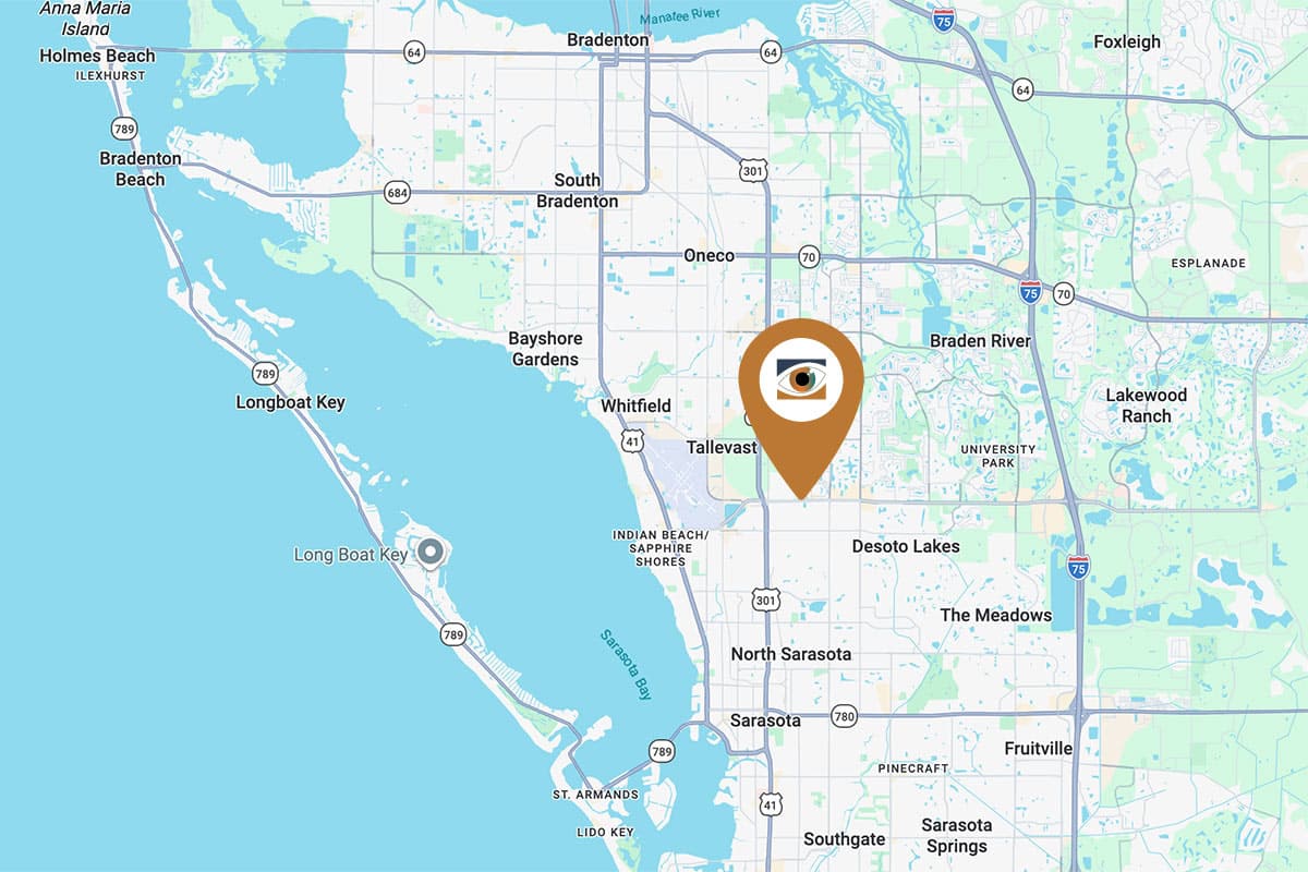
Sarasota - University Parkway Office
Located just west of I-75 on University Parkway in University Health Park.
Our University Parkway location serves patients from Sarasota, Lakewood Ranch, Bradenton, and surrounding areas. This office provides comprehensive evaluation and treatment for retinal vein occlusion, including ongoing monitoring and injection based therapies to manage retinal swelling and stabilize vision.
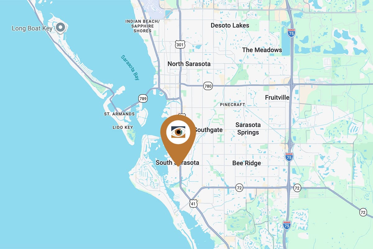
Sarasota - South Tamiami Trail Office
Located on South Tamiami Trail just south of Bee Ridge Road.
Our South Tamiami Trail location serves patients throughout Sarasota, Siesta Key, and nearby Sarasota County communities. This office offers prompt diagnosis and treatment for retinal vein occlusion, with a focus on protecting central vision and coordinating care with primary physicians when cardiovascular risk factors are involved.

Venice Retina Office
Located in central Venice near Venetia Bay Boulevard.
Patients in Venice, North Port, Englewood, and surrounding areas of southern Sarasota County can receive evaluation and treatment for retinal vein occlusion at this location. Our team provides individualized treatment plans designed to reduce retinal swelling and preserve vision over time.
Expert Retinal Vein Occlusion Treatment
At Shane Retina, we specialize in retinal vein occlusion treatment. Our team of experts has years of experience treating all types of retinal and macular conditions. We’ll find the right treatment for your needs and prevent further complications that could come as a result of the vein occlusion. Our thorough exam may involve dilation, blood tests, photographs, or other specialized imaging of the retina.
Contact Us At Shane Retina
There are effective ways to treat all forms of vein occlusion, so don’t delay your appointment if you are experiencing sudden, painless loss of vision in Sarasota, Bradenton, or Lakewood Ranch. If left untreated, vein occlusions can lead to glaucoma (intense eye pressure) and macular edema. Contact us today to get high-quality retinal vein occlusion treatment at one of our locations in Sarasota and Manatee County.
Get More Information About Retinal Vein Occlusion
For more information regarding this condition, check out our patient services education page. You can also visit the American Society of Retina Specialists resources about branch retinal vein occlusion or central retinal vein occlusion.